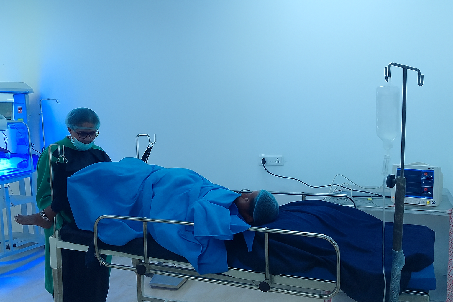
X-ray Basics
X-ray Basics is covered through structured sessions, demonstrations, discussion and applied examples suited to undergraduate medical learning.
Radiodiagnosis helps students see disease patterns and connect images with clinical findings.
The Department of Radiodiagnosis introduces students to X-ray, ultrasound, CT, MRI concepts, imaging anatomy, radiation safety and diagnostic interpretation.
At VRIMS, the department is planned as an active academic environment where students learn through lectures, demonstrations, tutorials, seminars, practical exposure and continuous feedback. The goal is to make every topic clinically meaningful and professionally useful.
Students are encouraged to connect basic concepts with real health needs, ethical practice and patient-centred communication.
Every area is taught with a balance of conceptual clarity, practical exposure and clinical relevance.
X-ray Basics is covered through structured sessions, demonstrations, discussion and applied examples suited to undergraduate medical learning.
Ultrasound is covered through structured sessions, demonstrations, discussion and applied examples suited to undergraduate medical learning.
CT Concepts is covered through structured sessions, demonstrations, discussion and applied examples suited to undergraduate medical learning.
MRI Concepts is covered through structured sessions, demonstrations, discussion and applied examples suited to undergraduate medical learning.
Imaging Anatomy is covered through structured sessions, demonstrations, discussion and applied examples suited to undergraduate medical learning.
Radiation Safety is covered through structured sessions, demonstrations, discussion and applied examples suited to undergraduate medical learning.
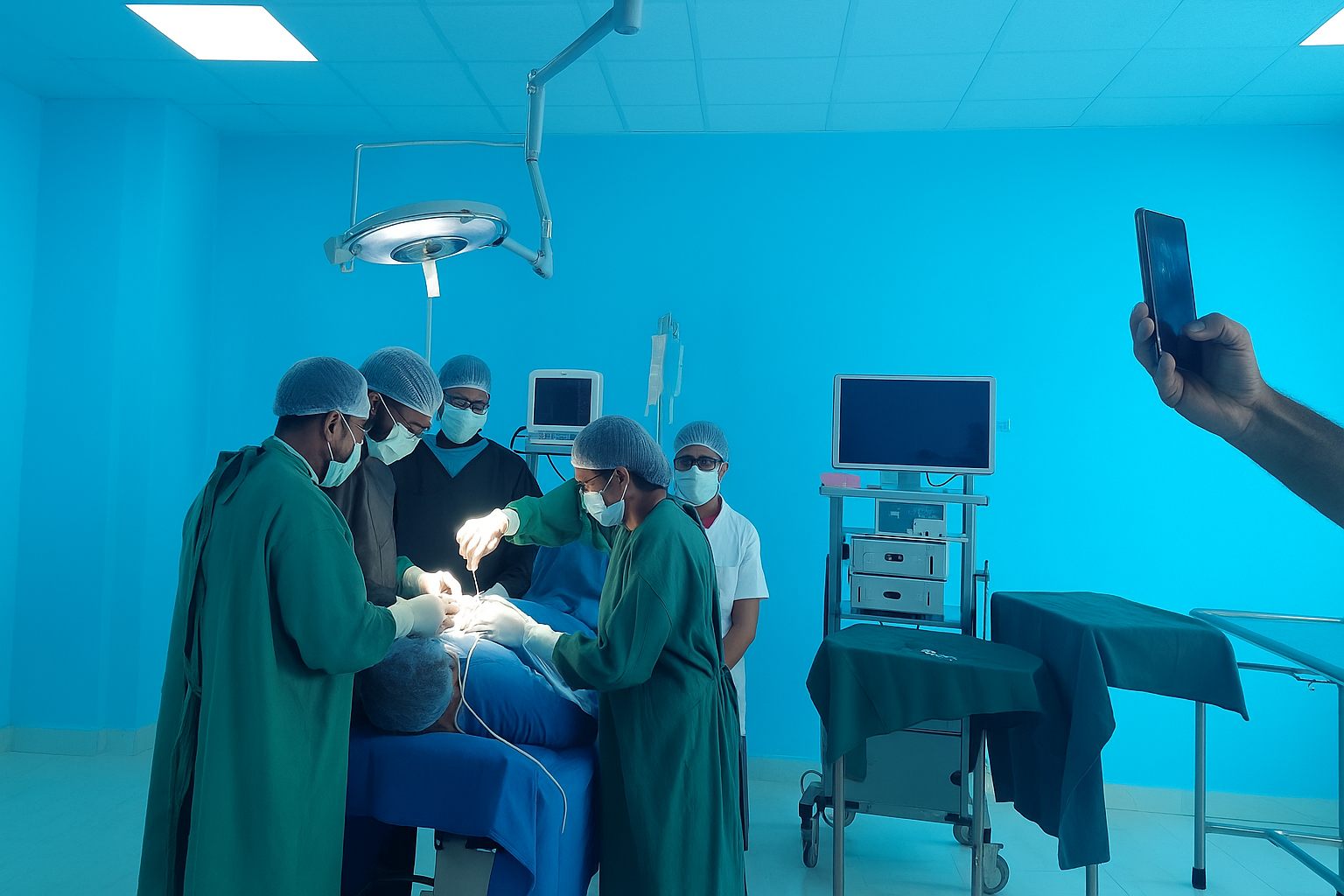
Regular lectures, small-group discussions and faculty-led academic sessions build a clear foundation.
Practical classes, demonstrations, clinical observation and skill-based learning connect theory with practice.
Topics are linked with other departments so students understand medicine as one connected discipline.
Seminars, viva practice, case presentation and feedback help students improve steadily through the course.
The department page is written to help students, parents and visitors understand how the subject is actually experienced at VRIMS.
X-ray Basics is approached through concept-building, faculty explanation, relevant examples and repeated academic reinforcement. Students are encouraged to ask questions, compare findings, understand terminology and connect the topic with patient care wherever applicable.
Ultrasound is approached through concept-building, faculty explanation, relevant examples and repeated academic reinforcement. Students are encouraged to ask questions, compare findings, understand terminology and connect the topic with patient care wherever applicable.
CT Concepts is approached through concept-building, faculty explanation, relevant examples and repeated academic reinforcement. Students are encouraged to ask questions, compare findings, understand terminology and connect the topic with patient care wherever applicable.
MRI Concepts is approached through concept-building, faculty explanation, relevant examples and repeated academic reinforcement. Students are encouraged to ask questions, compare findings, understand terminology and connect the topic with patient care wherever applicable.
Imaging Anatomy is approached through concept-building, faculty explanation, relevant examples and repeated academic reinforcement. Students are encouraged to ask questions, compare findings, understand terminology and connect the topic with patient care wherever applicable.
Radiation Safety is approached through concept-building, faculty explanation, relevant examples and repeated academic reinforcement. Students are encouraged to ask questions, compare findings, understand terminology and connect the topic with patient care wherever applicable.
Students are introduced to the scope of the subject, its relevance in MBBS and the professional expectations of departmental learning.
Core topics are explained through structured teaching, diagrams, demonstrations, tutorials and guided reading.
Practical or clinical sessions help learners observe real patterns, perform basic tasks and discuss applied examples.
Important concepts are linked with other departments so students can understand disease, diagnosis and care more completely.
Regular tests, viva preparation, assignments and feedback help students measure progress and strengthen weak areas.
The department supports students with learning resources, academic mentorship, discipline-specific exposure and an environment focused on professional growth.
Students receive academic support through approachable faculty, doubt-clearing sessions and guided learning.
Learning areas are used for demonstrations, practical exercises, procedural understanding and visual reinforcement.
The subject is connected with patient care, diagnostics, safety and responsible decision making wherever relevant.
Communication, documentation, teamwork, ethics and respect for patients are reinforced throughout training.
Beyond written notes, the department uses a mix of active learning formats so students stay engaged throughout the academic year.
Topic-wise teaching for conceptual clarity and exam readiness.
Small-group academic support for discussion and doubt clearing.
Visual and practical sessions that make difficult topics easier to understand.
Student presentations that build confidence, communication and subject command.
Clinical or applied examples to show why the subject matters in practice.
Written work and guided learning tasks to strengthen retention.
Regular academic guidance to help students improve steadily.
Focused recap sessions for important concepts and assessment preparation.
The department is not presented as a short subject note. It is shaped as a working academic unit where students understand the subject, practise its methods and learn why it matters in real medical care.
X-ray Basics is explained with definitions, diagrams, applied examples and repeated classroom reinforcement so students can move beyond memorisation and understand the logic of the subject.
Ultrasound is supported through practical demonstrations, visual teaching, observation, guided discussion and faculty feedback so learners can connect what they read with what they see.
CT Concepts helps students build clinical reasoning, compare normal and abnormal findings, understand important terminology and prepare for integrated learning with other departments.
Students are encouraged to maintain notes, attend sessions regularly, revise important topics, participate in seminars and discuss doubts early so learning remains steady throughout the academic year.
The department gives importance to correct academic language, clear presentation, careful recording of observations and respectful communication with faculty, peers, patients and attendants wherever applicable.
By combining theory, practical exposure, assessment, revision and mentorship, the department prepares students for university examinations, clinical postings, internship responsibilities and lifelong medical learning.
Every topic in Department of Radiodiagnosis is connected with observation, explanation, practice and professional behaviour, giving visitors a clearer view of how the department contributes to the VRIMS academic journey.
Develop a strong understanding of essential subject concepts and their role in medicine.
Build observation, examination, interpretation, documentation and problem-solving abilities.
Practise with discipline, empathy, scientific curiosity and respect for patient dignity.
Prepare for higher clinical learning, internship responsibilities and lifelong medical education.
Share your details and the admissions team will contact you.